Our Services
CT
Computed Tomography is an imaging method that uses X-ray, along with powerful computers, to generate high-resolution images of any part of the body.
To collect medical information about the brain, spine, chest, abdomen, and pelvis, CT scans are routinely used. It is also a highly efficient way of obtaining details of the skeletal system. 3-D images are produced to help diagnose in certain clinical cases. A CT scan can help identify the location and problem of a stroke or bleeding within the brain. This can also be used for examining the sinuses and facial structure.
- Reduce the need for exploratory surgeries
- Determine when surgeries are necessary
- Accurate
Lowering the bar on radiation exposure
LDCT: Low Dose CT is a special type of Chest CT scan that screens the lungs for cancer. The USPSTF recommends annual screening for lung cancer with low-dose computed tomography (LDCT) in adults aged 55 to 80 years who have a 30 pack-year smoking history and currently smoke or have quit within the past 15 years.
All CT exams utilize dose control measures to keep the radiation dose as low as possible.
- Abdomen (Gallbladder, Liver, Pancreas, Kidneys)
- Nothing to eat or drink 4 hours prior to your exam. (You may drink water)
- You may be asked to pick up a Barium Drink to have the day before and day of your appointment.
- Pelvis
- Nothing to eat or drink 4 hours prior to your exam. (You may drink water)
- You may be asked to pick up a Barium Drink to have the day before and day of your appointment.
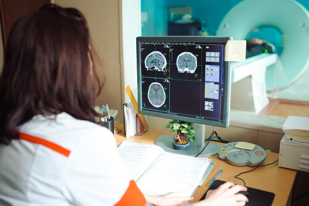

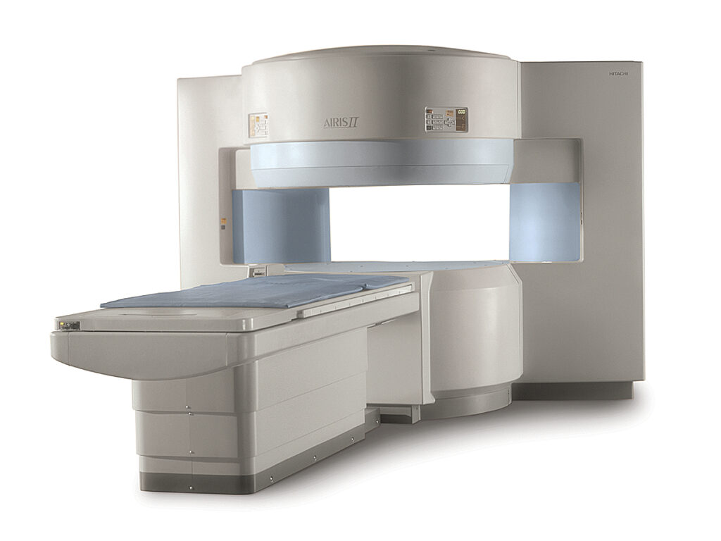
Open MRI (Magnetic resonance imaging)
Open MRIs use magnets to take images of the inside of a human body. Our open MRI machine, the Hitachi Airis II, is a reliable system that meets a variety of needs. Our Open MRI is perfect for patients who are anxious or have a phobia with small spaces. Our open design provides a more relaxed and comfortable experience. Under these controlled circumstances, our radiologists trained in neuroradiology, body imaging and orthopedic radiology can interpret your scans.
Before undergoing an Open MRI, alert your doctor if you have implants, metal clips, or other metallic objects in your body as these can be affected by the magnet.
- Spacious
- Comfortable
- Easy
- Abdomen (Gallbladder, Liver, Pancreas, Kidneys) or MRCP
- Nothing to eat or drink 6-8 hours prior to your exam. (You may drink water)
- Pelvis
- Nothing to eat or drink 4 hours prior to your exam. You may be asked to empty your bladder prior to starting the study.
- Any MRI with contrast injection (Brain, IAC’s, Orbits, Pituitary, Extremities, Spine)
- Nothing to eat or drink 4 hours prior to your exam. (You may drink water)
- Abdomen (Gallbladder, Liver, Pancreas, Kidneys) or MRCP
US
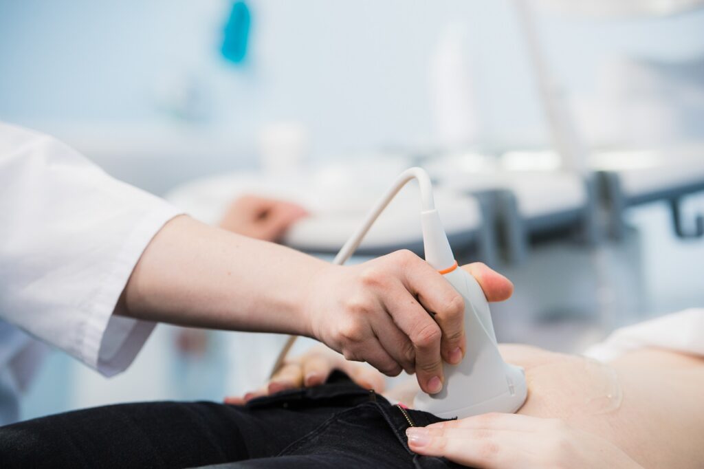
Ultrasound is a diagnostic tool that uses a transducer, or probe, to generate sound waves and computer technology to produce pictures of internal structures. Our ultrasound services cover a wide range of applications, from thyroid, to venous, vascular, scrotum, hip, cranial, abdomen, and many more. Ultrasound can be used to evaluate breast lumps and abnormalities seen on mammograms.
Using a special form of ultrasound called a Doppler, the speed and direction of flowing blood can be measured and illustrated in color pictures. This Doppler technique allows radiologists to find blocked blood vessels.
Ultrasound is safe, painless and can often provide information that eliminates the need for more expensive tests or surgery.
- Safe
- Quick
- Easy
- Aorta, kidney, or upper abdominal studies:
- Do not eat or drink for eight hours prior to the procedure.
- Pregnancy, bladder, pelvic, pelvic with transvaginal:
- Empty your bladder 90 minutes prior to the study and drink 32 ounces of water. You can take up to 30 minutes to finish the entire 32 ounces of water; the fluid must be consumed one hour prior to your appointment time. Do not empty your bladder until the exam is complete.
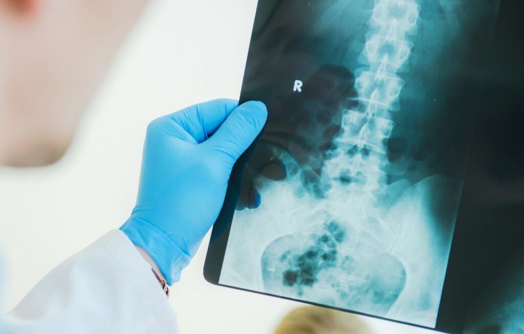
X-Ray
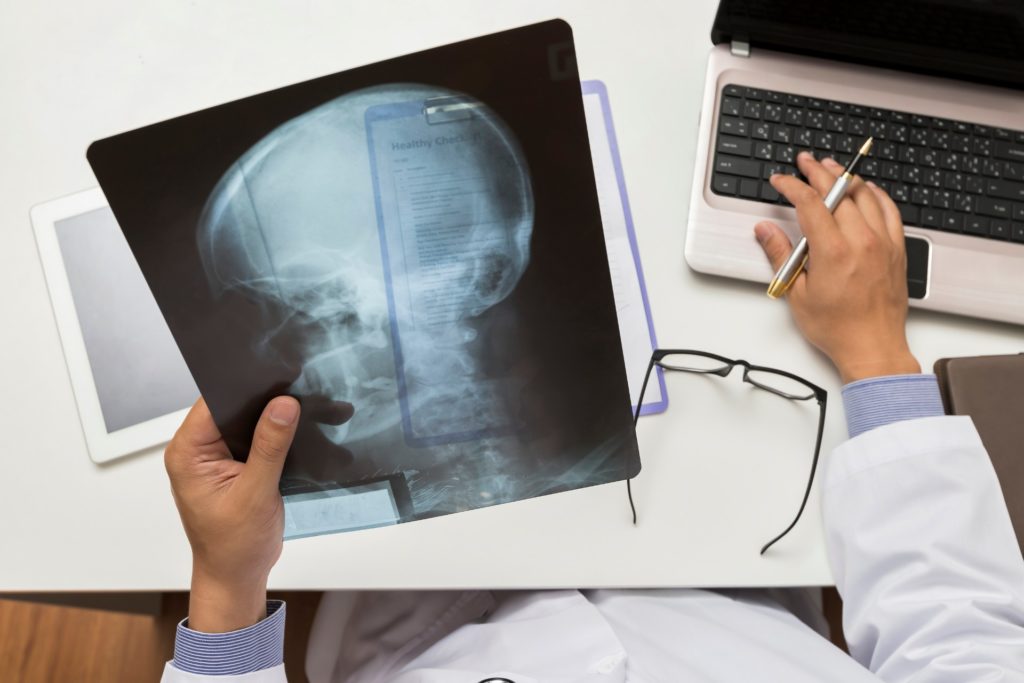
Digital X-rays have countless advantages over conventional methods of imaging. While traditional X-rays have generally been considered safe, digital X-rays use 80 percent less radiation than traditional images.
X-rays are forms of electromagnetic radiation similar to light. They are higher in energy and reach the body so to acquire images of inner structures. X-rays are used to expose a photographic plate or digital sensor. X-rays show portions of the body in different gray colors, with certain tissues appearing whiter (e.g. bone) than organs (e.g. lungs) that appear darker on the image.
- Painless
- Noninvasive
- Quick
DEXA Scans
DEXA scans are completed to help you better understand your bones’ density. This X-Ray scan determines your bones’ health and risk of fracture due to osteoporosis.
- Bone Density Testing
- Preventitive
- Simple